TARGET PROJECT 4
The eye as a window into sickle cell disease morbidity
Project Lead: Jing Jin, M.D.

Dr. Jin is an Assistant Professor of Ophthalmology and Pediatrics, Sidney Kimmel Medical College at Thomas Jefferson University. She earned her Doctor of Medicine degree at West China University of Medical Sciences, Chengdu, China, her Doctor of Philosophy at Department of Physiology, Boston University, Boston. Dr, Jin completed her residency and fellowship from Duke University of Medical Center, Durham, NC. Her Ph.D. training in basic visual science and clinical training in pediatric ophthalmology have prepared me to serve as Principal Investigator for the proposed study.
She is currently an Ophthalmologist, Division of Ophthalmology, Department of Surgery at Nemours Children’s Health DE, and is a member of the American Association for Pediatric Ophthalmology and Strabismus. Dr. Jin has provided care for children with eye disease for over 20 years. She has been using Optical Coherence Tomography (OCT) to examine the long-term retinal changes in pediatric eye diseases. One of the diseases she has focused on is sickle cell disease (SCD). Dr. Jin’s expertise in the clinical care of children and her experience performing clinical and basic science research have prepared her well to serve as principal investigator of this proposal.
Project Summary/Abstract:
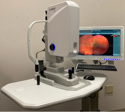
Sickle cell disease (SCD) is the most common genetic mutation among the US population, affecting approximately 100,000 Americans. As a systemic illness, SCD attacks multiple organs including the eyes. Although SCD can affect nearly all structures in the eye, sickle cell retinopathy (SCR) is the vision threatening complication with a reported 10% blindness rate. SCR damage often begins in childhood and progresses with age. Early in SCR, small peripheral retinal vascular occlusions, also known as “retinal stroke”, are typically asymptomatic. However, as the disease progresses, abnormal blood vessels begin to grow (neovascularization or proliferative change). Advanced proliferative SCR can lead to intraocular hemorrhage, retinal detachment and vision loss. Currently, there are no proven methods to prevent SCR, and treatment options are focused on deterring the progression of proliferative SCR. Prompt diagnosis of SCR is an important step for early treatment, which can prevent vision loss. The diagnostic strategies focus on visualizing signs of retinal stroke and abnormal vessel growth. NIH guidelines recommend annual screening for SCR by dilated funduscopic examination of the eye beginning at age ten, which has low sensitivity. The pathological process of retinal neovascularization is a complex phenomenon under active investigation. The research proposed in this application, The Eye as a Window into Sickle Cell Disease Morbidity, will utilize a combination of standard and novel noninvasive diagnostic modalities to examine retinal circulation and structure changes in children with SCD. Using these techniques will improve our ability to accurately diagnose and monitor SCR in children and provide a better understanding of the mechanism of the disease. Furthermore, the value of sensitive and specific assessments of ocular changes in SCD is not limited to saving vision. For many neurologic conditions and systemic illnesses, ocular findings aid in both diagnosis and monitoring of overall disease progression. Thus, findings from the proposed ophthalmologic examinations may elucidate a link between retinal damage and damage to other at-risk organs, especially the brain in SCD. Through this study, we will refine our ability to predict progression, assess therapeutic efficacy, develop more effective monitoring guidelines and initiate early interventions. Correlating retinal findings with clinical data may provide insight into other vascular complications of SCD, such as cerebrovascular disease. The long-term goal of this study is to enhance early detection of SCR and develop treatment strategies to slow the progression of SCR toward proliferative retinopathy and blindness, lowering the societal burden of SCR and improving the quality of life for these patients.

Publications:
https://www.ncbi.nlm.nih.gov/myncbi/1vGNDoEnZ6hs75/bibliography/public/